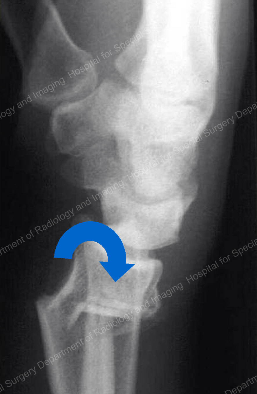

The Holstein-Lewis fracture thus has an association with radial nerve palsy (22 %). At this particular level, the radial nerve courses distally through the lateral intermuscular septum and is in direct contact with the adjacent bone, leaving the nerve susceptible to injury. The injury is a spiral fracture of the distal third of the humerus, typically as a consequence of blunt trauma, with the distal fragment displaced such that the proximal aspect is deviated radially. The Holstein-Lewis fracture was first described by American orthopedic surgeons Arthur Holstein and Gwilym Lewis. Surgical techniques to address recurrent anterior instability include labral repair or bone reconstruction if the degree of bone loss is severe. These lesions can be diagnosed with a CT (especially 3-D reconstructions) or MRI. Similar to Hill-Sachs, this lesion may result in anterior shoulder joint instability and recurrent dislocations. Any concern for this injury warrants non-emergent magnetic resonance imaging (MRI), which provides evaluation for labral as well as osseous injury. The inferior glenoid rim should be closely interrogated on pre- and post-reduction shoulder radiographs. An osseous or bony Bankart fracture is a chip fracture of the anterior inferior glenoid cortical rim on which the labrum rests (Fig. The soft tissue Bankart lesion is an injury to the anterior or anteroinferior glenoid labrum, the fibrocartilagenous structure that surrounds and deepens the bony glenoid. Named after the renowned English orthopedic surgeon Arthur Bankart, the Bankart lesion is often associated with the Hill-Sachs lesion due to their common mechanism of injury. We also briefly review the mechanism of each injury, associated complications, any follow-up imaging needed, and treatment. We illustrate fundamental descriptors of each injury that a clinician should expect in a radiology report. In this two-part series, our goal is to provide emergency providers with consistent, accurate definitions and depictions of commonly and less frequently encountered extremity fracture eponyms, keying in on important imaging features that differentiate these fractures.

Unfortunately, the imprecise use of eponyms can result in confusion and miscommunication. Eponymous extremity fractures are commonly encountered in the emergency setting and are frequently used in interactions amongst radiologists, emergency clinicians, and orthopedists. Their use allows physicians to quickly provide a concise description of a complex injury pattern. Eponyms are embedded throughout medicine they can be found in medical literature, textbooks, and even mass media.


 0 kommentar(er)
